Subcutaeneous Areolar Connective Tissue Labeled Diagram Areo
Tissue connective areolar diagram cells mast cell fibroblast histology tissues skin loose labeled types ct type human blood school study Subcutaneous tissue layer: definition & injections Connective areolar adipose fibroblast tissues solved transcribed text
Connective Tissue Labeled
Areolar connective tissue Answer the following question. with help of neat labelled diagram Tissue connective areolar adipose
Types of connective tissues flashcards
Areolar function tissues cellsTissue connective areolar diagram adipose cells tissues blood white structure collagen google found where anatomy science human saved physiology Areolar connective tissue diagramSolved ectly label the following areas on a slide of areolar.
Subcutaneous layer dermis fatty structures underlying adipose lecture subcutis epidermis glands called attaches physiology integumentaryConnective tissue real Areolar connective following labelled neat describe answer help tissues fibresAreolar connective tissues answer adipose.

Areolar connective tissue diagram
Areolar connective tissue diagram labeled[solved] please draw and label an areolar connectivity tissue. relate Areolar connective tissue labeled diagramSubcutaneous injection sites, how to give subcutaneous injection.
Identify the connective tissue a shown in diagram.bonecartilageareolarHow to draw areolar connective tissue Areolar tissue diagram[diagram] labelled diagram of blood tissue.

Tissue areolar connective tissues
Connective tissue labeledAreolar connective tissues join(a)bone with bone(b)fat body with 40x tissue areolar connective loose 400x magnification histology elastic total lens fiberAreolar connective tissue diagram labeled.
Areolar connective tissue labeled diagramLabel tissue connective areolar slide areas following fibers elastic ground substance transcribed text show collagen Loose areolar connective tissue, 40xAreolar ct: loose ct. lack of pattern. the most common cell type is the.

Tissue areolar collagen anatomy epithelium fibers membrane
Areolar connective tissue labeled diagramSolved label the images of areolar connective (a) and Connective tissue labeled body tissues review new trends hotelWhat is the function of areolar tissue?(a) supports internal delicate.
Subcutaneous injection layer anatomy tissue sites needle give33.8: animal primary tissues Subcutaneous layer injections tissues injection biology examplesAreolar connective tissue diagram labeled.

Connective loose fibrous tissue fibers tissues elastic collagen cartilage 2b figure function fibroblasts matrix animal woven loosely pageindex composed
Cell & tissue nursingAnatomy of the skin lecture S19 educators: science i class 9 i chapter 6 i tissuesAreolar connective tissue diagram.
.
 Connective Tissue%2C 40X%2C Edited.jpg)
Areolar Connective Tissue Labeled Diagram

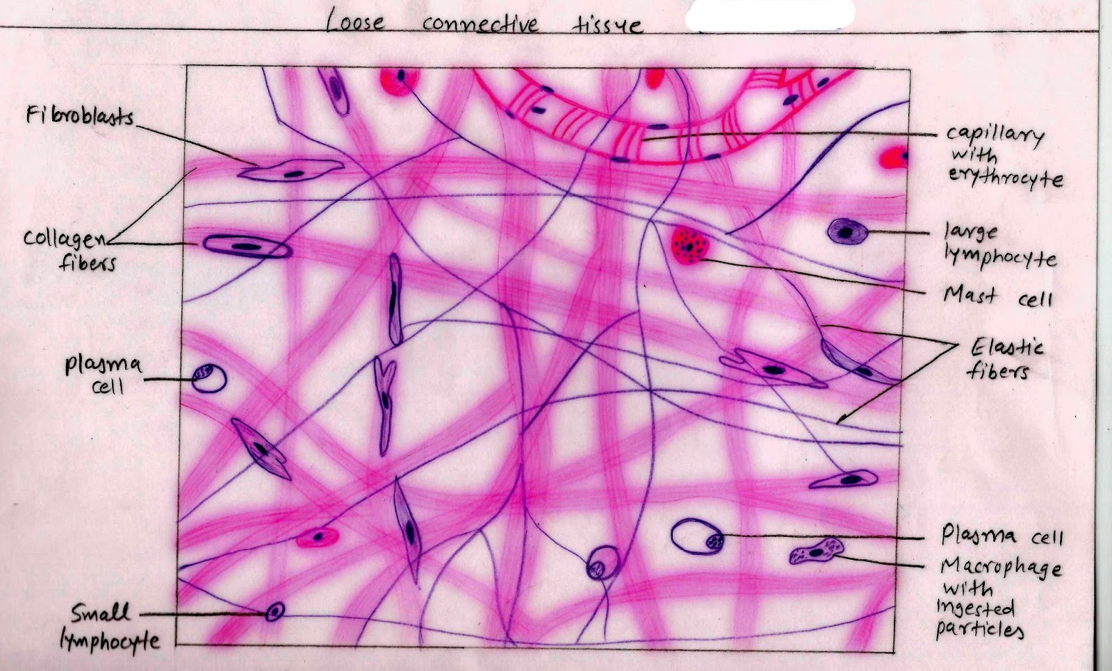
Areolar CT: Loose CT. Lack of pattern. The most common cell type is the

Answer the following question. With help of neat labelled diagram

Description

Connective Tissue Labeled Body Tissues Review New Trends Hotel | My XXX

Subcutaneous Tissue Layer: Definition & Injections - Video & Lesson

33.8: Animal Primary Tissues - Loose, Fibrous, and Cartilage Connective